Home

seau amplitude spontané myelin figures Faisabilité relais par exemple

Electron micrograph showing electron-dense laminated myelin figures... |  Download Scientific Diagram
Electron micrograph showing electron-dense laminated myelin figures... | Download Scientific Diagram

Morphology of Lyotropic Myelin Figures Stained with a Fluorescent Dye | The  Journal of Physical Chemistry B
Morphology of Lyotropic Myelin Figures Stained with a Fluorescent Dye | The Journal of Physical Chemistry B
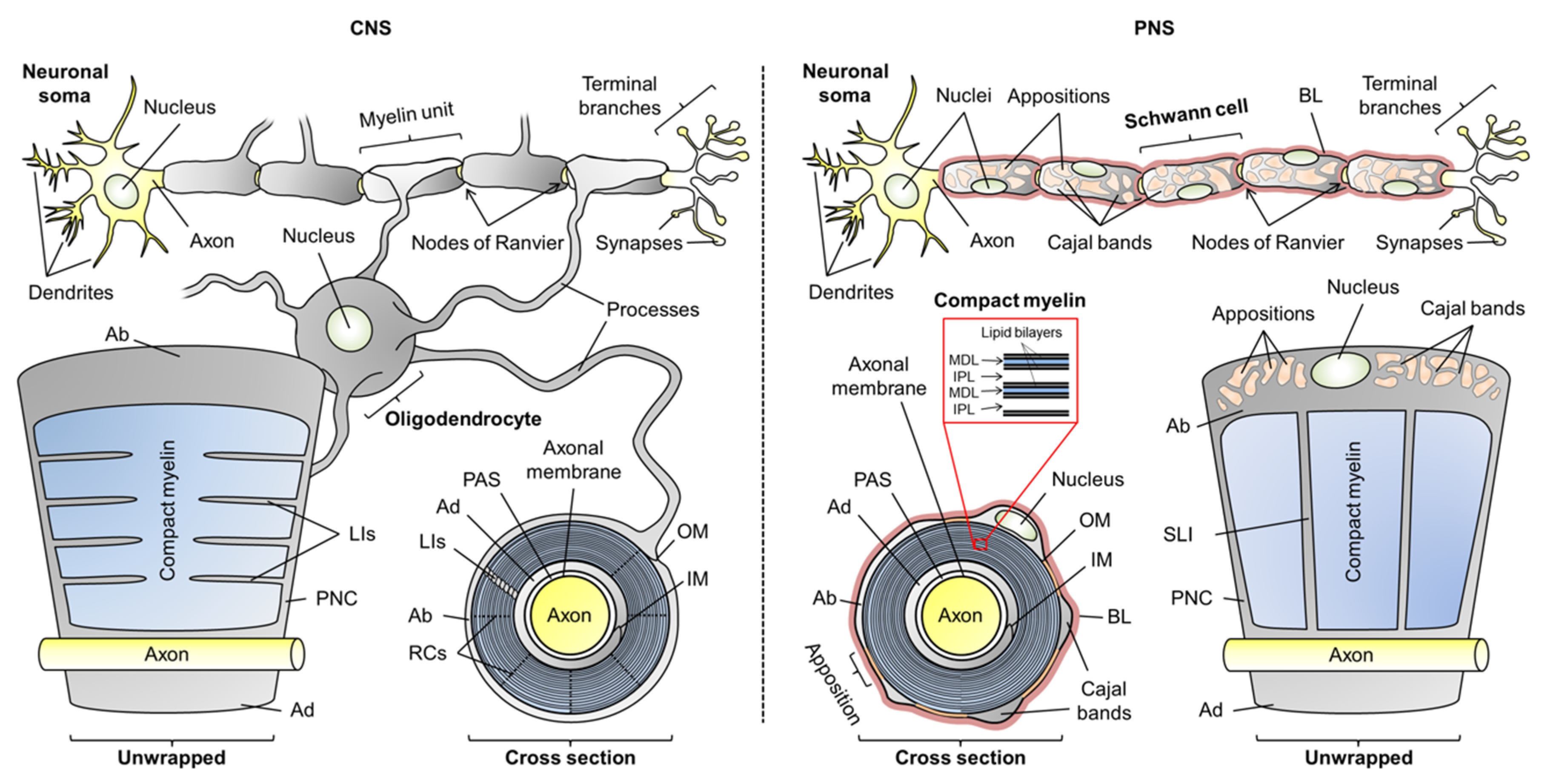
Figure 4-10 from Myelin Formation, Structure and Biochemistry | Semantic  Scholar
Figure 4-10 from Myelin Formation, Structure and Biochemistry | Semantic Scholar

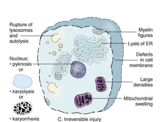
Cell injury | PPT
Cell injury | PPT

Cells | Free Full-Text | Flexible Players within the Sheaths: The  Intrinsically Disordered Proteins of Myelin in Health and Disease
Cells | Free Full-Text | Flexible Players within the Sheaths: The Intrinsically Disordered Proteins of Myelin in Health and Disease

Synthetic myelin figures immobilized in polymer gels - Soft Matter (RSC  Publishing)
Synthetic myelin figures immobilized in polymer gels - Soft Matter (RSC Publishing)

Analysis of oxidative processes and of myelin figures formation before and  after the loss of mitochondrial transmembrane potential during  7β-hydroxycholesterol and 7-ketocholesterol-induced apoptosis: comparison  with various pro-apoptotic chemicals ...
Analysis of oxidative processes and of myelin figures formation before and after the loss of mitochondrial transmembrane potential during 7β-hydroxycholesterol and 7-ketocholesterol-induced apoptosis: comparison with various pro-apoptotic chemicals ...

Cell injury | PPT
Cell injury | PPT

Cells with myelin figures and lipid droplets | Wellcome Collection

myelin figure - www.medicoapps.org

Myelin figures associated with the cytoplasmic membranes in human  medulloblastoma
Myelin figures associated with the cytoplasmic membranes in human medulloblastoma

Cell injury part 1 | PPT

Figure 5. | Mechanisms of Primary Axonal Damage in a Viral Model of  Multiple Sclerosis | Journal of Neuroscience
Figure 5. | Mechanisms of Primary Axonal Damage in a Viral Model of Multiple Sclerosis | Journal of Neuroscience
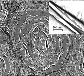
Myelin figures in the cytoplasm of notochordal cell; Pb exposure;... |  Download Scientific Diagram
Myelin figures in the cytoplasm of notochordal cell; Pb exposure;... | Download Scientific Diagram

SGU SOM - T4 - Cell Injury and Adaptation II Flashcards | Quizlet

Myelin figures in the basal-granulated cells of human Brunner's glands. |  Semantic Scholar
Myelin figures in the basal-granulated cells of human Brunner's glands. | Semantic Scholar

Cell Injury and Cell Death - NEET PG Pathology

Synthetic myelin figures immobilized in polymer gels - Soft Matter (RSC  Publishing) DOI:10.1039/B701455D
Synthetic myelin figures immobilized in polymer gels - Soft Matter (RSC Publishing) DOI:10.1039/B701455D

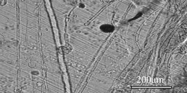
Formation of myelin figures in a typical contact experiment. A lump of... |  Download Scientific Diagram
Formation of myelin figures in a typical contact experiment. A lump of... | Download Scientific Diagram
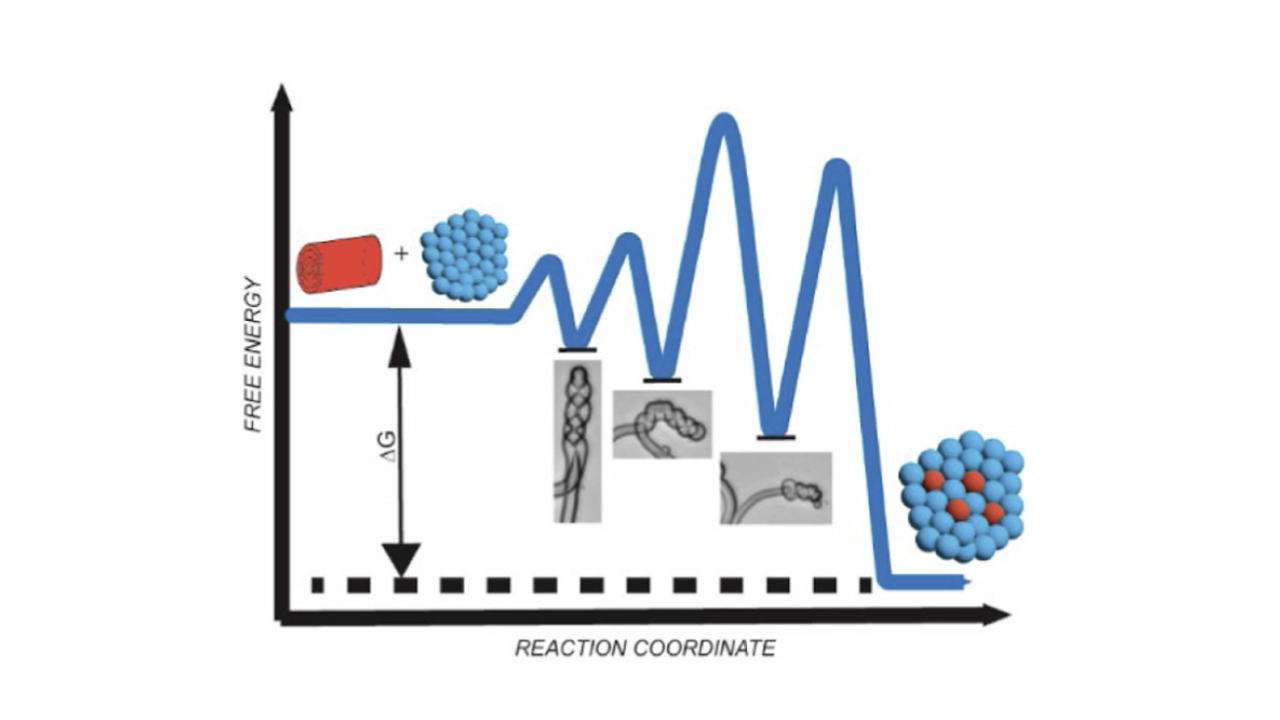
Surfactant-Mediated Solubilization of Myelin Figures: A Multistep  Morphological Cascade | Parikh Lab | parikhpeople
Surfactant-Mediated Solubilization of Myelin Figures: A Multistep Morphological Cascade | Parikh Lab | parikhpeople

Uncoupling of Myelin Assembly and Schwann Cell Differentiation by  Transgenic Overexpression of Peripheral Myelin Protein 22 | Journal of  Neuroscience
Uncoupling of Myelin Assembly and Schwann Cell Differentiation by Transgenic Overexpression of Peripheral Myelin Protein 22 | Journal of Neuroscience

Figures
Figures

Growth of Myelin Figures from Parent Multilamellar Vesicles | Langmuir

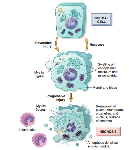
Pathologic Cell Injury and Cell Death II – Necrosis – The Art Of Medicine

Figure 9 from In addition to their well-recognized function in the  phagocytosis and digestion of foreign materials which enter their  environment, alveolar macrophages may also participate in the turnover of  surfactant, as
Figure 9 from In addition to their well-recognized function in the phagocytosis and digestion of foreign materials which enter their environment, alveolar macrophages may also participate in the turnover of surfactant, as

Notas de un Cientifico - Myelin figures The high lipid content of the red  cell envelope is expressed morphologically by the appearance of  microspherules and myelin figures in damaged cells. Myelin figures
Notas de un Cientifico - Myelin figures The high lipid content of the red cell envelope is expressed morphologically by the appearance of microspherules and myelin figures in damaged cells. Myelin figures